


Summary
Vanderbilt researchers have developed a system for enhancing the imaging capabilities of optical coherence tomography (OCT), a tool commonly used to monitor and treat patients with retinal disease. The image resolution of OCT, however, is intrinsically limited. Ideally, a contrast agent could be used to highlight specific parts of the retina within the image, but dye alone is largely ineffective because of the way OCT generates the image. Photothermal heating solves this problem by creating local zones of tissue expansion which can be distinctly detected by OCT. Photothermal-OCT is safe, effective, and enhances the imaging power of a tool widely used by opticians.
Addressed Need
Careful monitoring of optical diseases such as macular degeneration, diabetic retinopathy, and glaucoma is vital to patient well-being. OCT is a non-invasive imaging tool which produces a 3D image of the retina, allowing the clinician to visualize disease progression both inside and out of the surgical environment. However, image resolution is intrinsically limited in OCT making it difficult to highlight those areas of the retina of clinical interest. Vanderbilt researchers have created an imaging device which combines OCT with photothermal heating to enhance image specificity, which is safe and effective.
Technology Description
Photothermal-OCT is performed with the use of contrast agents, typically a dye which is safe for use in eyes. Once injected, the pigment is retained in the target tissue. A photothermal light source applies thermal energy, at a specified frequency, causing a localized expansion of the dye-containing tissue. These changes are detected and translated to the image so that the tissue stands out and can be monitored. The present approach can also be used with endogenous absorbers, such as melanin.
Commercial Applications
Photothermal-OCT is ideal for intraoperative imaging in conjunction with OCT machinery. OCT imaging is also used in other clinical settings for monitoring optical disease progression, and the present approach provides additional contrast and specificity to these existing applications.
Intellectual Property Status
Issued US patent: 10,682,053
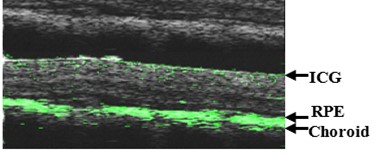
Figure 1: OCT image (grey) overlayed with a photothermal-OCT image (green) of an animal retina with contrast agent.